Lower Brain Levels of mGluR5 Receptor Found in Fragile X Men
Written by |
Brain scans in men with fragile X syndrome showed reduced levels of the mGluR5 receptor — a protein receptor known to help regulate nerve cell communication — compared with healthy men, a pilot study found.
These findings may help explain the failure of previous clinical trials to ease the symptoms of the genetic disorder, which is characterized by developmental delays and social and behavioral problems.
The study, “Reduced Expression of Cerebral Metabotropic Glutamate Receptor Subtype 5 in Men with Fragile X Syndrome,” was published in the journal Brain Sciences.
Fragile X is caused by mutations in the FMR1 gene, which provides instructions for the protein FMRP. This protein can be found in many tissues, including the brain, where it controls the production of several proteins at synapses — sites where nerve cells communicate.
The loss of FMRP causes an increase in protein production and excitatory signaling in the brain, meaning there’s a greater likelihood of activating a nerve cell. This would normally be mediated by a protein receptor called metabotropic glutamate receptor 5, or mGluR5.
Studies suggest that the mGluR5 receptor is found at higher levels in the brain of people with fragile X, meaning that there’s too much of the protein receptor. However, assessments in the living human brain are still lacking.
Past research has focused on targeting mGluR5 as a potential treatment approach in fragile X, but no significant benefits have been seen in clinical trials. However, recent evidence showed the altered availability of mGluR5 at the cell membrane, where it interacts with other molecules, which may have caused the failure of the clinical studies.
As such, measuring mGluR5 levels in the living brains of men with fragile X may help understand these past failures and provide needed information for future clinical trial design.
To this end, investigators based at the Institute for Neurodegenerative Disorders (IND), in Connecticut, and The Johns Hopkins University (JHU), in Maryland, designed a study to directly measure the density and distribution of mGluR5 in the brains of fragile X patients.
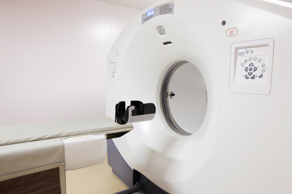
The team used MRI and PET (positron emission tomography) scans to trace a radioactive molecule called FPEB that binds to the receptor.
One of the study’s main goals was to determine the safety and feasibility of a testing procedure that included MRI and PET scans in people with fragile X, who generally have behavioral control issues. This was accomplished by advanced training in a mock scanner, and with the help of practice sessions using behavioral psychology as well as assistance from trained parents.
One patient at the JHU site had never had a PET scan before, and had met with a psychologist to prepare him for previous MRIs. According to the researchers, the man’s mother worked with the investigators to prepare him for the scans.
“His mother began practicing relaxation and holding still while listening to MRI sounds for weeks before the session,” the researchers wrote.
At the IND site, seven men with fragile X — ranging in age from 23 to 34 years — were assessed, along with three men with typical development who were included as controls. In turn, data from JHU included the assessments of two patients, ages 24 and 27, and five age-matched healthy people (controls).
The results from IND showed no significant differences in mGluR5 levels between patients and controls. In contrast, the JHU site findings found lower mGluR5 levels in men with fragile X than in the controls.
By combining the results from both groups, the researchers found that mGluR5 was significantly reduced in eight areas of the brain in fragile X men as compared with controls. These areas were the anterior cingulate, caudate, occipital, parietal, posterior cingulate, putamen, temporal, and thalamus, which are involved in functions such as learning, memory, movement, emotions, and sensory processing.
“We showed that the proposed protocol of MR and PET scans in one day is feasible in individuals with FXS [fragile X syndrome] who have received mock scanner training by an experienced behavioral psychology team,” the researchers wrote.
“Most importantly, we found for the first time that quantified mGluR5 expression using [FPEB] is reduced in the living human brain of men with FXS [fragile X] in contrast to healthy normal age- and sex-matched controls,” they wrote.
“Larger studies with additional molecular biomarkers are needed to expand on the feasibility finding of this protocol,” the investigators added, to further assess whether it may help “the design of clinical trials of glutamatergic agents in FXS.”
In addition, future studies will benefit from using the same protocols at different sites as a way to improve the consistency of results, the investigators said.