
White Matter Lesions Among Brain MRI Pattern of FXTAS, Case Report Describes


A new case report described a characteristic imaging pattern of fragile X‑associated tremor/ataxia syndrome (FXTAS) in a 63-year-old man, which included white matter lesions in a brain region called the cerebellum.
The study, “MRI findings in fragile X‑associated tremor/ataxia syndrome,” was published in the journal Acta Neurologica Belgica.
FXTAS, a common condition in families affected by fragile X syndrome, is a late-onset disorder, usually appearing after age 50, and characterized by movement and cognitive problems. While people with fragile X syndrome have over 200 CGG repeats in the FMR1 gene, those with FXTAS have a “premutation” in the same gene that consists of 55 to 200 such repeats. The C in CGG stands for cytosine, while the G is for guanine; these are two of the four building blocks of DNA.
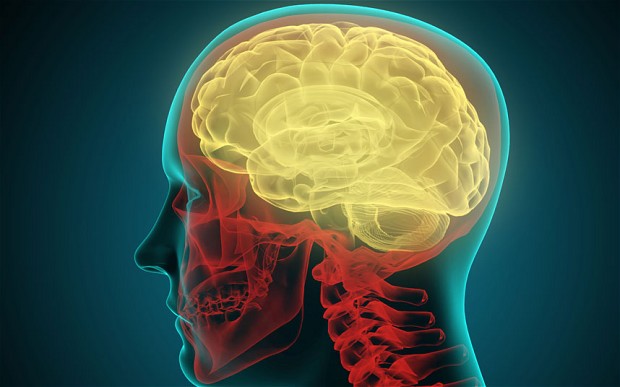
In people with FXTAS, magnetic resonance imaging (MRI) of the brain usually shows damage in the cerebellum — a key region that controls movement and balance — and in the white matter, which is composed of nerve fibers that transmit signals between different areas of the brain.
The team at University Hospitals Leuven in Belgium described the case of a man with 108 CGG-repeats in FMR1. His symptoms included action tremors during voluntary movement, postural tremors — such as when holding the arms outstretched — and a slight intention tremor. Intention tremors are produced with purposeful movement toward a target, such as lifting a finger to touch the nose.
To ease these tremors, the patient was started on primidone, an anti-seizure medication which works by decreasing abnormal electrical activity in the brain.
The man also experienced a subtle decrease in coordination of the extremities, called appendicular ataxia, decreased lower limb reflexes, difficulty on tandem gait — when the toes of the back foot touch the heel of the front foot at each step — and erectile dysfunction. He did not have any cognitive complaints.
A brain MRI showed the characteristic signs of FXTAS.
MRI images, or “sequences,” can be T1, T2, or FLAIR — Fluid Attenuated Inversion Recovery — depending on how certain tissues look. For example, on T1, the white matter appears light and the cerebrospinal fluid (CSF), which is the liquid surrounding the brain and spinal cord, appears dark. Meanwhile, on T2, white matter appears dark gray and CSF bright. FLAIR sequences are similar to T2, but the CSF appears dark.
This patient showed the middle cerebellar peduncles (MCP) sign, which is typical in FXTAS. It consists of white matter hyperintensities — meaning lesions show up with increased brightness on the MRI — in the middle cerebellar peduncles, which connect the cerebellum to the brain stem.
Other findings were hyperintensity in the splenium of the corpus callosum, a thick nerve tract connecting the cerebral hemispheres — the splenium being its thickest part. He also showed mild hyperintensity in the white matter surrounding the cerebral ventricles, or the cavities where CSF circulates.
Thinning in the corpus callosum and generalized cerebral atrophy, or shrinkage, also were found.
“The presence of intention tremor (a major clinical diagnostic sign) and the MCP sign (a major radiological diagnostic sign) along with other radiologic signs, were consistent with the diagnosis of FXTAS,” the researchers said.